Restriction Fragment Length Polymorphism (RFLP) analysis, a veritable molecular fingerprinting technique, once reigned supreme in the realm of genetic diagnostics and identification. Imagine it as a molecular sculptor, chiseling away at the vast genomic landscape to reveal the unique signatures etched within an individual’s DNA. This technique, while partially eclipsed by more modern methodologies, still holds historical significance and offers valuable insights into the intricate world of inherited traits and disease susceptibility.
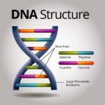
At its core, RFLP leverages the power of restriction enzymes – molecular scalpels that recognize and cleave DNA at specific nucleotide sequences. These sequences, known as restriction sites, are not uniformly distributed throughout the genome. Their presence or absence, and the distances between them, vary from individual to individual due to subtle genetic variations known as polymorphisms. These polymorphisms, like hidden geological strata, dictate the precise lengths of the DNA fragments generated by restriction enzyme digestion.
The RFLP procedure unfolds in a series of meticulously orchestrated steps.
- DNA Extraction: The journey begins with the isolation of DNA from a biological sample, be it blood, tissue, or even ancient bone fragments. This is the raw material upon which the molecular sculpting will take place.
- Restriction Digestion: Next, the extracted DNA is incubated with a specific restriction enzyme. This enzyme diligently scans the DNA molecule, seeking out its cognate restriction sites and cleaving the DNA at these precise locations. The result is a collection of DNA fragments of varying lengths.
- Gel Electrophoresis: These fragments are then separated by size using gel electrophoresis, a technique that exploits the inherent negative charge of DNA. When subjected to an electric field, the fragments migrate through a gel matrix, with smaller fragments traversing more rapidly than larger ones. This process effectively sorts the fragments by length, creating a distinct banding pattern.
- Southern Blotting: The separated DNA fragments within the gel are then transferred to a nitrocellulose or nylon membrane via a process known as Southern blotting. This transfer immobilizes the DNA, making it accessible for subsequent hybridization. Think of it as lifting a delicate fingerprint from a fragile surface.
- Hybridization: The membrane is then incubated with a labeled DNA probe – a short, single-stranded DNA sequence that is complementary to a specific region of interest within the genome. This probe, carrying a radioactive or fluorescent tag, seeks out and binds to its complementary sequence on the membrane.
- Detection: Finally, the location of the hybridized probe is visualized using autoradiography (for radioactive probes) or fluorescence detection. This reveals the specific DNA fragments that contain the sequence recognized by the probe, creating a distinctive RFLP pattern.
The clinical applications of RFLP analysis are diverse, spanning the realms of disease diagnosis, forensic science, and paternity testing. Consider its role in inherited disease diagnostics.
For diseases like Huntington’s disease or cystic fibrosis, where specific genetic mutations are known to cause the condition, RFLP can be used to determine whether an individual carries the mutated gene. By designing a probe that targets a region near the disease-causing gene, clinicians can analyze the RFLP patterns to identify individuals who are homozygous for the normal allele, heterozygous carriers of the mutation, or homozygous for the mutated allele, indicating the presence of the disease.
Furthermore, RFLP played a pivotal role in the early days of forensic science. The unique RFLP pattern of an individual’s DNA, much like a fingerprint, could be used to identify suspects in criminal investigations or to establish familial relationships in paternity disputes. The power of RFLP in this context stemmed from the immense variability in RFLP patterns across the population, making it a highly discriminating tool.
While RFLP analysis has been largely supplanted by more rapid and efficient techniques like PCR-based methods and next-generation sequencing, it remains a cornerstone of molecular biology history. Its inherent limitations, including the requirement for relatively large amounts of high-quality DNA and the labor-intensive nature of the procedure, paved the way for the development of newer, more user-friendly techniques.
The legacy of RFLP lies not only in its past applications but also in its contribution to our understanding of genomic variability and its impact on human health. It serves as a testament to the ingenuity of scientists who dared to dissect the intricate code of life, revealing the hidden genetic landscapes that shape our individuality and predispose us to disease. Though a veteran technique, its impact on the field of genomics reverberates even today, shaping the landscape of modern molecular diagnostics.
Modern applications do exist, although less common. In certain situations, particularly in resource-limited settings or for specific research purposes where the detailed analysis of larger genomic regions is desired, RFLP can still offer advantages. For example, it may be used in microbial typing to differentiate strains of bacteria or viruses based on variations in their genomic restriction patterns. Moreover, RFLP analysis can be valuable in analyzing complex genomic rearrangements or insertions, where the detection of large structural variations is crucial.
Despite its relative decline in clinical diagnostics, RFLP continues to serve as a valuable tool in certain specialized applications. The methodology stands as a symbol of the foundational work done in the realm of genetics and molecular biology, a testament to human ingenuity. It remains a vital piece of the puzzle in our ongoing quest to decipher the complexities of the genome.


Leave a Comment