
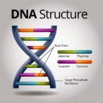
Deoxyribonucleic acid, or DNA, is the fundamental blueprint of life. The intricate double helix holds the genetic instructions for every organism, dictating everything from eye color to predisposition for certain diseases. But observing that double helix in a biology textbook or a science documentary is one thing; unlocking the secrets held within its sequences is quite another. DNA analysis has revolutionized fields from medicine to criminology, offering unprecedented insights into the very fabric of existence. The profound implications of DNA analysis stem from its ability to reveal not just who we are, but where we came from, and potentially, where we are going.
Several methodologies exist for deciphering the genetic code. Each technique offers unique strengths and is employed for specific applications. This exposition delves into the primary types of DNA analysis, elucidating their principles and applications.
1. Sequencing: Unveiling the Genetic Text
DNA sequencing is the process of determining the precise order of nucleotides (adenine, guanine, cytosine, and thymine) within a DNA molecule. This is the bedrock of modern genomics. It is perhaps the most fundamental technique in DNA analysis.
* Sanger Sequencing: Considered the “gold standard” for many years, Sanger sequencing, also known as chain-termination sequencing, involves synthesizing DNA strands of varying lengths, each terminating with a modified nucleotide. These fragments are then separated by size, and the sequence is read based on the terminal nucleotide of each fragment. Though reliable, Sanger sequencing is relatively slow and expensive for large-scale projects. It’s still useful for verifying results and sequencing shorter DNA fragments.
* Next-Generation Sequencing (NGS): NGS technologies have revolutionized DNA sequencing by allowing for massively parallel sequencing of millions or even billions of DNA fragments simultaneously. This dramatically increases throughput and reduces the cost per base. NGS platforms like Illumina, Ion Torrent, and PacBio utilize different chemistries and approaches, but all share the common goal of rapidly generating vast amounts of sequence data. Application areas are whole-genome sequencing, exome sequencing (focusing on protein-coding regions), and RNA sequencing (transcriptomics). NGS is crucial in personalized medicine, allowing for tailored treatments based on an individual’s genetic makeup. Furthermore, ecological studies leverage NGS to characterize microbial communities within complex environments.
2. Polymerase Chain Reaction (PCR): Amplifying the Signal
PCR is a technique used to amplify specific DNA sequences exponentially. This molecular photocopying enables researchers to generate millions or billions of copies of a target DNA region from a very small starting sample. This is a really powerful tool.
* Basic PCR: Involves repeated cycles of denaturation (separating the DNA strands), annealing (binding of primers to the target sequence), and extension (DNA synthesis by a polymerase enzyme). Primers are short, synthetic DNA sequences that flank the target region. The process amplifies only the region between the primers. This is broadly applied in diagnostics, forensics, and research. For instance, PCR is a cornerstone of COVID-19 testing.
* Quantitative PCR (qPCR): Also known as real-time PCR, qPCR allows for the quantification of DNA or RNA templates during the amplification process. Fluorescent dyes or probes are used to monitor the amount of amplified product in real-time. This technique is invaluable for measuring gene expression levels, detecting pathogens, and quantifying viral loads. The dynamic monitoring offers a precise assessment of the initial target quantity.
* Reverse Transcription PCR (RT-PCR) This technique is used to amplify RNA sequences. Reverse transcriptase enzyme converts RNA into complementary DNA (cDNA), which is then amplified by PCR. RT-PCR is useful for studying gene expression and identifying RNA viruses. It combines the specificity of PCR with the ability to analyze RNA.
3. Fragment Analysis: Measuring DNA Size
Fragment analysis techniques determine the size and quantity of DNA fragments. These methods are often used to detect variations in DNA sequences that affect fragment length.
* Capillary Electrophoresis: This is a high-resolution technique used to separate DNA fragments based on their size and charge. Fluorescently labeled DNA fragments are injected into a narrow capillary, and an electric field is applied. The fragments migrate through the capillary at different rates depending on their size. A detector measures the fluorescence of each fragment as it passes by. This allows for precise determination of fragment size and quantity. Capillary electrophoresis is widely used in microsatellite analysis for parentage testing and forensic identification. It is also used for analyzing PCR products and detecting mutations.
* Restriction Fragment Length Polymorphism (RFLP): Although largely supplanted by more modern techniques, RFLP involves digesting DNA with restriction enzymes, which cut DNA at specific sequences. Variations in these restriction sites between individuals result in different fragment lengths. These fragments are then separated by gel electrophoresis and visualized. RFLP was one of the earliest methods for DNA fingerprinting and is still used in some applications.
4. Hybridization-Based Assays: Finding Specific Sequences
Hybridization assays rely on the principle that complementary DNA strands will bind to each other under specific conditions. This allows for the detection of specific DNA sequences within a sample.
* Microarrays: Microarrays consist of a large number of DNA probes attached to a solid surface. A sample of DNA or RNA is labeled and hybridized to the microarray. The probes that bind to the labeled sample are detected, allowing for the simultaneous analysis of thousands of genes or DNA sequences. Microarrays are used in gene expression profiling, SNP genotyping, and comparative genomic hybridization. This is a comprehensive approach to analyzing genetic variation.
* Fluorescence In Situ Hybridization (FISH): FISH involves hybridizing fluorescently labeled DNA probes to chromosomes or DNA sequences within cells or tissues. This allows for the visualization of specific DNA sequences within their natural context. FISH is used in cytogenetics to detect chromosomal abnormalities, such as deletions, duplications, and translocations. It is also used in cancer diagnostics to identify specific gene amplifications or deletions. This offers a spatial perspective on genetic alterations.
5. Emerging Technologies: The Cutting Edge
The field of DNA analysis is constantly evolving, with new technologies emerging all the time.
* CRISPR-Based Diagnostics: CRISPR (Clustered Regularly Interspaced Short Palindromic Repeats) technology, primarily known for gene editing, is also being adapted for diagnostic applications. CRISPR-based diagnostics utilize the specificity of CRISPR systems to detect the presence of specific DNA or RNA sequences. These assays can be highly sensitive and specific, offering a rapid and accurate means of detecting pathogens or genetic mutations. Its adaptability and speed are particularly promising.
* Nanopore Sequencing: Nanopore sequencing involves passing DNA molecules through tiny pores. Changes in electrical current as the DNA passes through the pore are used to identify the sequence of nucleotides. Nanopore sequencing offers the potential for long-read sequencing, which can be particularly useful for resolving complex genomic regions. Its portability and real-time capabilities are also advantageous. This can provide a more complete picture of the genome.
These diverse DNA analysis techniques represent a powerful arsenal for unlocking the secrets of the genome. Each method offers unique advantages, contributing to advancements across various scientific disciplines. The continued development and refinement of these techniques will undoubtedly lead to even greater insights into the complexities of life.


Leave a Comment